http://www.pinkmonkey.com/studyguides/subjects/biology-edited/chap14/14_13.gif
http://www.slideshare.net/mrskennedy/protist-notes
http://www.coleharbourhigh.ednet.ns.ca/library/images/bio11m21.gif
http://www.coleharbourhigh.ednet.ns.ca/library/images/bio11m22.jpg
http://www.biosci.ohio-state.edu/~eeob/new/webgallery3/images/amoeba.jpg
http://www.microscopy-uk.org.uk/mag/imgdec02/paramecium.jpg
http://nyms.org/Gallery/diatoms.jpg
http://www.gpmatthews.nildram.co.uk/microplants/spyrogyra002.jpg
http://www.designswan.com/wp-content/uploads/2008/slime_mold/slime_mold2.jpg
http://www.biology.neu.edu/images/redalgae.jpg
http://www1.istockphoto.com/file_thumbview_approve/1439011/2/istockphoto_1439011_brown_algae_seaweed_specimen.jpg
http://128.128.175.244/fedora/get/data:26015/LocalImage
http://www.microscopy-uk.org.uk/mag/indexmag.html?http://www.microscopy-uk.org.uk/mag/wimsmall/smal1.html
http://www.pclaunches.com/entry_images/0207/26/bacteria-dna.jpg
http://nanopedia.case.edu/image/water%20flea.jpg
http://campus.queens.edu/faculty/jannr/images/critters/ostracods.jpg
http://hudsonregional.org/mosquito/images/copepod.jpg
http://www.microscopy-uk.org.uk/mag/smallimag/cosci3.jpg
http://www.microscopy-uk.org.uk/index.html?http://www.microscopy-uk.org.uk/ponddip/index.html
http://www.planet-pets.com/amoeba1.jpg
http://192.171.153.213/sci_programmes/Water/Lake%20Ecosystems/images/clip_image002.jpg
http://www.urbanrivers.org/web_images/diatoms.gif
http://www.mediterranea.org/cae/divulgac/peces/hydra.jpg
http://starcentral.mbl.edu/msr/rawdata/viewable/euglenatripteris_wbw.jpg
http://botit.botany.wisc.edu/Interface/web-lessons/Diversity/Chlorophyta/images/Spirogyra.jpg
http://www.gpmatthews.nildram.co.uk/animalcules/cypris02.jpg
http://www.lima.ohio-state.edu/biology/images/volvox.jpg
http://www.btinternet.com/~stephen.durr/volvoxc.jpg
http://water.me.vccs.edu/courses/ENV108/clipart/rotifer.jpg
http://www.ucmp.berkeley.edu/phyla/rotifera/philodina.jpg
http://www.hometrainingtools.com/images/942/250x225/ms-desmid.jpg?1227081449
http://microscopy.fsu.edu/primer/techniques/hoffmangallery/images/stentor.jpg
http://www.susq-town.org/hamsher/004%20Structure%20of%20a%20paramecium.jpg
http://www.wasdarwinright.com/images/Vorticellafeeds.jpg
Sunday, January 18, 2009
Actinosphaerium (single-celled)
Their size ranges form 200 - 1000 µm
Where to find them : Planktonic and amongst plants (especially fine-leaved).
They have hair-like pseudopodia called axopodia (which are often stiffened) that radiate outwards. There are a number of smaller species e.g. in the genera Actinophrys and Acanthocystis.
Classification :Kingdom - Protoctista, Class - Heliozoa
** These remind me of dandilions for some reason.**
Vorticella (single-celled)
 Vorticella
The 'bell' is up to 150 µm, with stalk up to 1 mm.
You can find them attached to algae and plants etc.
Vorticella are one of the peritrich protozoa. Although solitary, they often occur in groups to form a tiny jelly-like mass just visible to the naked eye. Their stalks contract like a spring (they remind me of a slinky). Some similar types e.g. Epistylis (not contractile) and Carchesium (contractile) are true colonies with branching tree-like stalks.
Classification : Kingdom Protoctista, Phylum Ciliophora
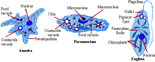
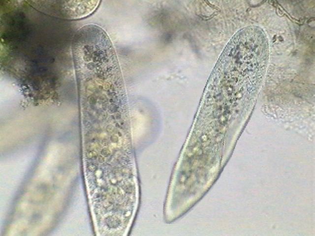
Paramecium (single-celled)

Their size range is between 60 - 300 µm.
You can find them amongst organic matter and Plankton.
They are one of the larger single-celled pond animals. Many protozoa are very small, but the larger Paramecium can just be seen as a speck swimming in pond water.
Classification :Kingdom Protoctista, Phylum Ciliophora
Stentor (single-celled)
 They can grow up to 2 - 3 mm long.
You can find these dandy creatures attached to plants/algae and planktonic.
They are one of the largest freshwater protozoans and larger than some multi-celled pond animals. When it is attached to a surface, it adopts the trumpet shape shown and the ring of cilia around the trumpet rim draw in water, together with the smaller organisms on which the Stentor feed. When the Stentor swims it changes to an oval shape.
The Stentor is often green in colour because of the algae (single-celled plants) associated with it. Like other large single celled creatures (e.g. amoebas) they have many nuclei. In Stentor you can see the nuclei as a 'string of pearls'. The large sphere is a water expelling vesicle. The cell is covered with tiny hair-like 'cilia'.
Classification :Kingdom Protoctista, Phylum Ciliophora
Desmid (single-celled)
 Desmids
Their Size varies greatly amongst speciesca 10 µm - 1 mm.
Where to find them : Planktonic, or on vegetation, particularly neutral to acidic waters.
They are an attractive and varied group of algae. And although the species vary widely in shape, they share the feature of being divided into two equal halves (semicells) which are often mirror images. A distinct constriction between the two semicells or gap in the plastids is usually seen.
Classification :Kingdom - Protoctista, Phylum - Gamophyta
** They look like little leeches to me.. GROSS!**
Rotifers (multicellular)
 Philodina
 Rotifers
there is a wide range amongst species size but around 25 µm - 1 mm.
Some species are planktonic, others are attached to plants, other organisms, stones or creeping on plants, mud surface etc.
Older books call them wheel animalcules. The head has a crown of cilia (the 'wheel organ'), which in some species looks as if it is rotating like a wheel.
Rotifer species show a wonderful variety of forms. Some move and contract like a leech (e.g. Philodina), others build 'houses'.
Rotifer species show a wonderful variety of forms. Some move and contract like a leech (e.g. Philodina), others build 'houses'.
Classification :Kingdom - Animalia, Phylum - Rotifera
Subscribe to:
Comments (Atom)